
Class 12 Biology Chapter 3 Human Reproduction The answer to each chapter is provided in the list so that you can easily browse through different chapters Assam Board HS 2nd Year Biology Chapter 3 Human Reproduction Question Answer.
Class 12 Biology Chapter 3 Human Reproduction


Also, you can read the SCERT book online in these sections Solutions by Expert Teachers as per SCERT (CBSE) Book guidelines. These solutions are part of SCERT All Subject Solutions. Here we have given Assam Board Class 12 Biology Chapter 3 Human Reproduction Solutions for All Subjects, You can practice these here.
(F). Long Answer Question : (Five Marks Each):
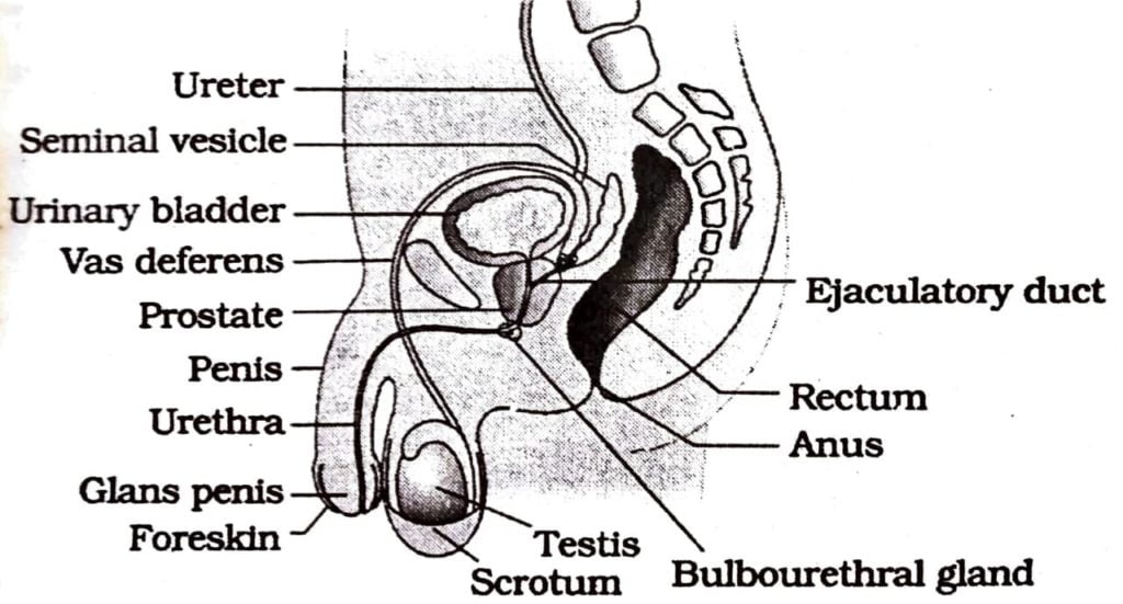
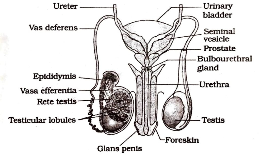
Q.1. Describe the structure of testes with neat labeled diagram.
Ans : The testes are situated outside the abdominal cavity within a pouch called scrotum. The scrotum helps in maintaining the low temperature of the testis necessary for spermatogenesis. In adults each testis is oval in shape, with a length of about 4 to 5 cm and a width of above 2 to 3 cm. The testis is covered by a dense covering. Each testis has about 250 compartments called testicular lobules.
Each lobule contains one to three highly coiled seminiferous tubules in which sperms are produced. Each seminiferous tubule is lined on its inside by two types of cells called male germ cells amd sertoli cells. The male germ cells undergo meiotic division and form sperm while sertoli cells provide nutrition to the germ cells. The interstitial space present in the outside region of the seminiferous tubule contain blood vessels and leydig cells.
Q.2. Describe the female reproductive system in human.
Ans : The female reproductive system consists of a pair of ovaries along with a pair of oviducts, uterus, cervix, vagina and the external genitalia located in pelvic region. These parts of the system along with a pair of mammary glands are integrated structurally and functionally to support the processes of ovulation, fertilization, pregnancy, birth and child care.
Ovaries are the primary female sex organs that produce the female gametes and several steroid hormones. The ovaries are located on each side of the lower abdomen. Each ovary is about 2-4 cm in length and is
connected to epithelium wall and uterus by ligaments. Each ovary is covered by a thin epithelium which encloses the ovarian stroma and it is divided into two zones a peripheral cortex and an inner medulla.
The oviduct or fallopian tube, uterus and vagina constitute the female accessary ducts. Each fallopian tube is about 10-12 cm long and extends from the periphery of each ovary to the uterus. The part closer to the ovary is funnel shaped infundibulum, the edges of which possess fingers like projections called fimbriae while help in collection of ovum after ovulation. The infundibulum leads to an ampulla and the last part of the oviduct is isthmus. The narrow lemon of the isthmus joins the oviduct.
The uterus is also known as womb which is like an inverted pear. It is supported by ligaments attached to the pelvic wall. The uterus opens into vagina through a narrow cervix.
The female external genitalia include mons pubis, labia majora, labia minora, hymen and clitoris.
The diagrammatic view of the female reproductive system is shown below :


Q.3. Give the structure of the mammary gland with neat diagram.
Ans : The characteristics of all female mammals is a functional mammary gland. The glands are paired structures that contain glandular tissue and variable amount of fat. The glandular tissue of each breast is divided into 15-20 mammary lobes containing clusters of cells called alveoli which secrete milk that is stored in the cavities of alveoli. The ovary opens into mammary tubules. The tubules of each lobe join to form a mammary duct. Several mammary ducts join to form a wider mammary ampulla which is connected to lactiferous duct through which milk is sucked out.

Q.4. Explain the process of spermatogenesis.
Ans : The process by which sperms are produced in the testes is known as spermatogenesis. It begins at the puberty. The spermatogonia present on the inside wall of seminiferous tubules multiply by mitotic divition and increase number. The spermatogonium is diploid. Some of the spermatogonia called primary spermatocyte periodically undergo meiosis and after completion of first meiotic division two equal haploid cells known as secondary spermatocytes. The secondary spermatocyte undergo second meiotic division to produce four equal haploid spermatids. The spermatids are transformed into spermatozoa or sperms by the process cells spermiogenesis.
Q.5. Describe the role of hormones in initiating spermatogenesis at puberty.
Ans : Spermatogenesis starts at the age of puberty due to significant increase in the secretion of gonadotropin releasing hormone (GNRH). It is a hypothalamic hormone. The increased levels of GnRH hormone acts at the anterior pituitary gland and stimulates secretion of two gonadotropins – Luteinizing hormone (LH) and follicle stimulating hormone (FSH) LH acts at the leydig cells and stimulates synthesis and secretion of androgens. Androgen in turn stimulates and process ofs FSH acts to help in the process of spermatogenesis.
Q.6. Describe the process of development of human embryo since one month of pregnancy.
Ans : Human pregnancy lasts 9 months and during this period of time the embryo undergoes different developmental stages. After one months of pregnancy, the embryo heart is formed. By the end of the second month, the foetus develops limbs and digits. By the end 12 weeks (first trimester), most of the major organ systems are formed like limbs and external genital organs are well developed. The first movement of the foetus and appearance of hair on the head are usually observed during fifth month. By the end of 24 weeks (second trimester), the body is covered with fine hair, eye lids separate and eyelashes are formed. By the end of nine months of pregnancy the fetus is fully developed and is ready to delivery.

Give One Word Technical Term : (Special One Mark Questions)
Q.1. The process by which spermatids are transformed into spermatozoa.
Ans : Spermiogenesis.
Q.2. The fluid filled cavity of the ovarian follicle.
Ans : Antrum.
Q.3. The membrane surrounds the secondary ocyte.
Ans : Zona pellucida.
Q.4. The first mensuration which begins at puberty.
Ans : Menarche.
Q.5. The daughter cells formed by cleavage.
Ans : Blastomere.
Q.6. The milk produced during initial few days of lactation.
Ans :Colostrum.
Q.7. Discuss the process of fertilization in human.
Ans : Fertilization in human involves the fusion of haploid male and female gamete to form diploid zygote. Fertilization in human beings is internal and takes place in the proximal part of the fallopian tube of the female. Fertilization involves the following processes-
(a) Approach of sperm to ovum : During the copulation, male insert his erectile penis in the vagina of female and releases about 3.5 ml of seminal fluid. It is called ejaculation. Seminal fluid contains around 200- 300 million sperms. The acidity of the vagina kills most of the sperms and only 100 sperm can reach the fallopian tube. The sperms can swim in the seminal fluid with lashing movement at the rate of 1-4 mm/minute. The sperms physiologically getting matured in the female genital tract which is called capacitation. It takes about 5-6 hours.
Ovum released from the Graafian follicle of ovary on 15th day of the menstrual cycle and the process is called ovulation. Ovum is traped by the fimbriae of the ampulla of fallopian tube. Ovum moves in the tube towards the uterus by peristalsis. At the time of ovulation egg is at secondary oocyte stage.
The fertilizability of human sperm is the female genital tract is of 12-24 hrs while the survival value is upto three days. Fertilizer liability period of ovum is only 24 hrs though it can live for 72 hrs.
To thin out the number of sperms, the ovum secretes a chemical substance, called fertilizin which has a spermophillic site-where sperms of species specific type can be found by their antifertilizin site. The fertilizin antifertilizin reaction is highly specific and to stop the polyspermy.
(b) Penetration of sperm : The sperm generally comes in contact (9) with ovum in the animal pole while the opposite site of ovum is called vegetative pole. Ovulation in human female occurs at secondary oocyte stage in which meiosis – I has been completed and first polar body has been released. Penetration of sperm is a chemical mechanism. Here, acrosome of sperm undergoes acrosomal reaction and releases sperm lysins which dissolve the egg envelop locally and make a passage for the penetration of sperm. Sperm lysins are acidic protein which contain an enzyme hyaluronidase. Only the sperm nucleus and middle piece enter the ovum. The tail is lost. In human, there is always monospermy.
(c) Cortical reaction: The penetration of sperm into the egg in a series of process where the egg membrane prevents the polyspermy. Sperm penetration into ovum also includes the following metabolic activities.
(i) The rate of protein synthesis increases.
(ii) The rate of respiration also increases.
(iii) The permeability of plasma membrane also increases.
(d) Fusion of gametic nuclei : The sperm entry stimulates the secondary oocyte to undergo meiotic II division which produces the ovum and second polar body. Inside the ovum, the sperm nucleus takes a definite path called copulation path. The centriole of the middle piece form a spindle. The nuclear membrane of the gametic nuclei degenerates and two sets of chromosomes initially lie on two poles and then mix up and this process is called amphimixis. The fertilised egg is new called zygote.
Q.8. Describe pregnancy and embryonic development in human.
Ans : After the fertilization, the fertilized egg undergoes a series of changes that taking place in side the female reproductive tract to form the embryo and this stage is called pregnancy. The pregnancy is mainly maintained by the progesterone hormone, so it is called pregnancy hormone. The embryonic development during pregnancy is discussed bellow:
(i) By the end of one month : Embryo’s heart starts to beat (4th week) and heart sound can be listened. Fore and hind limb develops. Optic vesicles are established. Liper and pancreatic bud is formed.
(ii) By the end of second month : Limbs become paddle like (5th week). Knees and elbows become distinct (8th week). Epidermis become single layered. Fusiform stomach is found with coiled intestine. Metanephros kidneys are formed. Brain, vesicle is formed.
(iii) By the end of third month : Epidermis multilayered, intestinal coil is found, islets of langerhans developed in pancreas, thymus, thyroid and parathyroid developed. External genitalia and limbs well developed. Prostate gland formation starts. Cerebellum and corpus callosum formed.
(iv) By the end of 4th month : Medullated nerve fibre formation begins.
(v) By the end of 5th month : First movement of foetus is found. Eruption of hair on head begin, Vagina canalised.
(vi) By the end of Sixth month : Fine hair on whole body is found. Separation of eye lids and fermentation of eye lashes takes place.
(vii) By the end of Eighth month : Testes descends in the scrotal sacs.
(viii) By the end of Ninth month : Fetus is fully developed and ready for birth.

Hi, I’m Dev Kirtonia, Founder & CEO of Dev Library. A website that provides all SCERT, NCERT 3 to 12, and BA, B.com, B.Sc, and Computer Science with Post Graduate Notes & Suggestions, Novel, eBooks, Biography, Quotes, Study Materials, and more.
