
Class 11 Biology Chapter 21 Neural Control and Coordination, AHSEC Class 11 Biology Question Answer, HS 1st year Biology notes to each chapter are provided in the list so that you can easily browse throughout different chapters Assam Board Class 11 Biology Chapter 21 Neural Control and Coordination Question Answer and select needs one.
Class 11 Biology Chapter 21 Neural Control and Coordination


Also, you can read the SCERT book online in these sections Solutions by Expert Teachers as per SCERT (CBSE) Book guidelines. These solutions are part of SCERT All Subject Solutions. Here we have given Assam Board Class 11 Biology Chapter 21 Neural Control and Coordination Solutions for All Subjects, You can practice these here.
Neural Control and Coordination
Chapter – 21
VERY SHORT ANSWER TYPE QUESTIONS
Q.1. Define Coordination?
Ans :- A process through which 2 or more organs interact and complement the functions of one an other is termed as coordination.
Q.2. Write the function of neurons.
Ans :- It can detect, receive and transmit different kinds of stimuli.
Q.3. What is somatic neural system.
Ans :- Somatic neural system relays impulses from the CNS to skeletal muscles.
Q.4. What is autonomic neural system?
Ans :- ANS transmit impulses from CNS to the involuntary organs and smooth muscles of the body.
Q.5. How ANS is classified?
Ans :- ANS is classified into sympathetic neural system and parasympathetic neural system.
Q.6. What is nodes of Ranvier.
Ans :- The gaps between 2 adjacent myelin sheath are called nodes of Ranvier.
Q.7. How synapse is formed?
Ans :- Synapse is formed by the membranes of a pre synaptic neuron and a post synaptic neuron, which may or may not be separated by a gap called synaptic cleft.
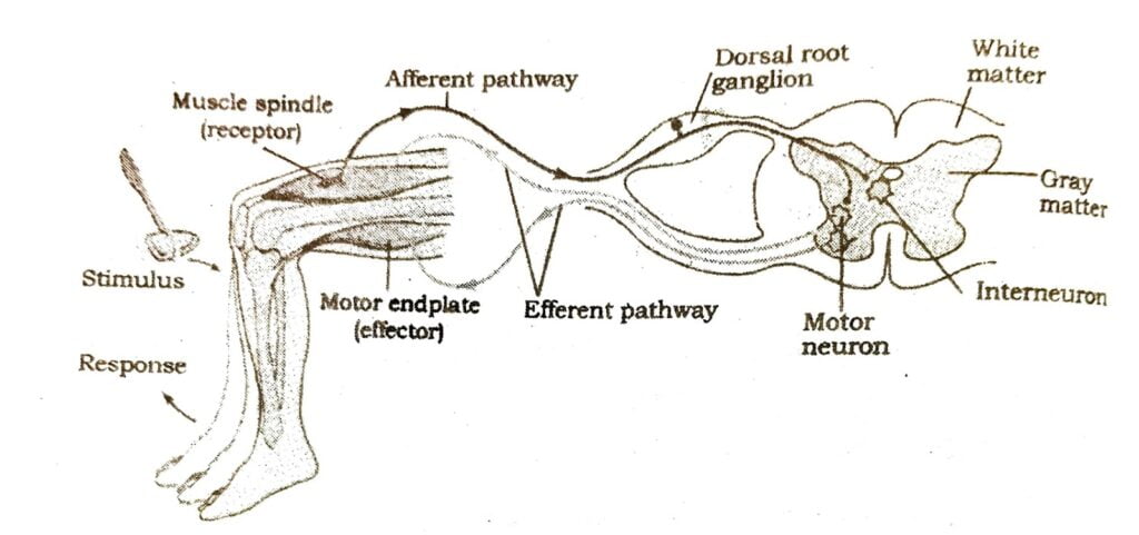
Q.8. Define reflex action.
Ans :- Actions which are controlled by the spinal cord is called reflex action.
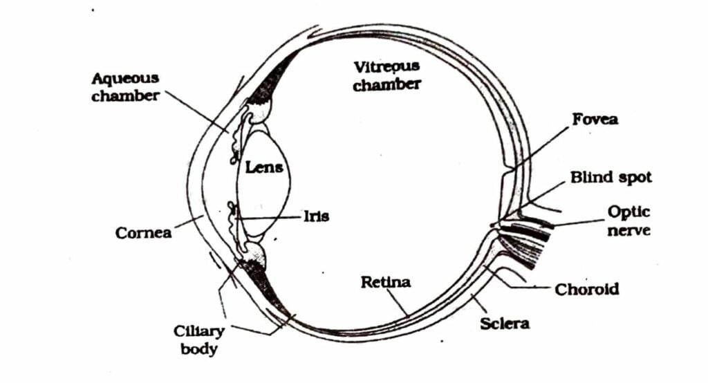
Q.9. Name the 3 layers of the eye?
Ans :- Sclera, Choroid and retina.
Q.10. Name the 3 layers of cells found in retention.
Ans :- Ganglion cells, bipolar cells and photoreceptor cells.
Q.11. What is blind spot?
Ans :- Region where photoreceptor cells are not present is termed as blind spot.
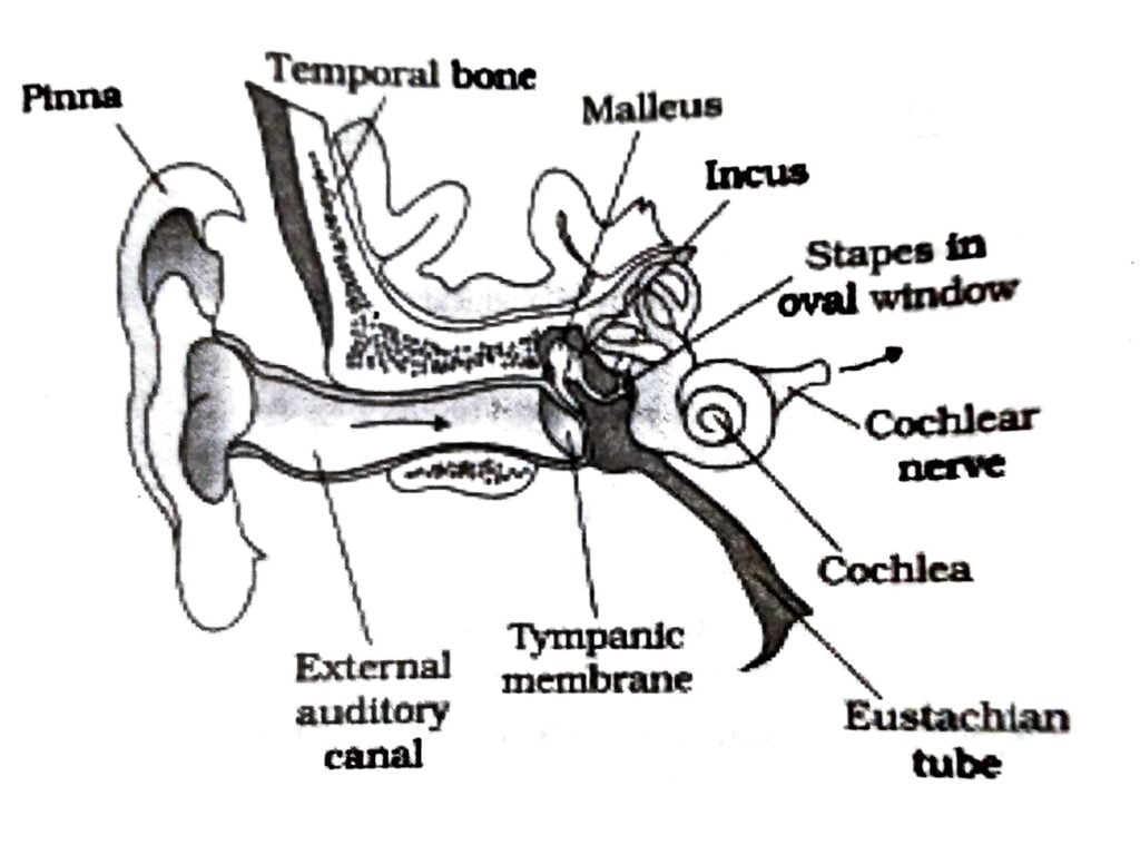
Q.12. What is cochlea?
Ans :- The coiled portion of labyrinth is called the cochlea.
Q.13. Mention 1 function of Eustachian tube?
Ans :- Eustachian tube helps in equaling the pressures on either sides of the ear drum.
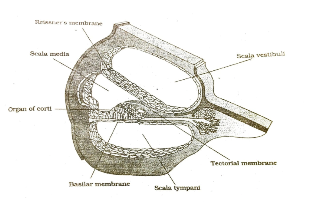
Q.14. What is organ of corti?
Ans :- Organ of Cortis a structure located on the basilar membrane which contain hair cells that act as auditory receptors.
Read Also: Camphor – Health Benefits, Uses, Qualities, and Side Effects
SHORT ANSWER TYPE QUESTIONS
Q.16. How neuron is classified based on the number of axon and dendrites.
Ans :- It is classified into 3 types. They are –
(i) Multipolar (1 axon and 2 or more dendrite).
(ii) Bipolar (1 axon and 1 dendrite).
(iii) Unipolar (Cell body with one axon only).
Q.17. What is neurotransmitter? Which part of neuron possess neurotransmitter.
Ans :- Neurotransmitters are chemical which help in transferring impulse through the synapse.
Synaptic knob of the axon possess synaptic vesicles where neurotransmitters are present.
18. Name the 2 types of synapses. In which type of synapses, transmission of impulse is faster.
Ans :- Electrical synapses and chemical synapses.
Impulse transmission across an electrical synapse is always faster than across a chemical synapse.
Q.19. What is cerebral cortex? Where is associations areas present? What is its function.
Ans :- Layer of cells which covers the cerebral hemisphere is called cerebra cortex.
In cerebral cortex, large region is there that are neither sensory nor motor in function called association areas. It is concerned with complex functions like intercessory association, memory and communication.
Q.20. Which part of the brain contain hypothalamus? What is its function?
Ans :- Fore brain contain hypothalamus.
Hypothalamus control body temperature, urge for eating and drinking. It also secretes hypothalamic hormones.
Q.21. Name the 2 photoreceptor cells present in our eye? How they are useful.
Ans :- Rods and cones. These cells contain light sensitive protein. Daylight vision and colour vision are the function of cones and the twilight vision is the function of rods.
Q.22. Where are myelinated and non climatic nerve fibres found.
Ans :- Myelinated Nerve fibres – In spinal cord and cranial nerves.
Unmyelinated nerve fibres – In autonomous and the somatic neural system.
Q.23. What is cranial meninges? Name the 3 layers present in it.
Ans :- The brain is covered by a layer just below the skull known as cranial meninges.
The three layers present in it are dura mater, arachnoid and pia mater.
Q.24. Give the diagrammatic presentation of reflex action showing knee jerk reflex.
Ans :-
Q.25. What is the difference between yellow spot on fovea and blind spot.
Ans :- Yellow spot cones for yellow pigment where maximum vision occurs whereas blind spot lack both rods and cones and optic nerve arises from this spot.
Q.26. Where are following parts located in our eye.
(a) Ciliary body.
(b) Iris.
(c) Aqueous humour.
(d) Pupil.
(e) Lens.
(f) Ganglion cell.
Ans :- (a) Ciliary body – Choroid layer.
(b) Iris – Choroids layer.
(c) Aqueous humour – Spece between the cornea and lens.
(d) Pupil – In choroid, in front of the lens, the aperture surrounded by the iris is called pupil.
(e) Lens – In choroid layer.
(f) Ganglion cell – In retina.
Q.27. Draw leveled diagram of our eye.
Ans :-

Q.28. Give a diagrammatic representation of the sectional view of cochlea and level the following parts. Organ of corti, scala tympani and Reissner’s membrane.
Ans :-
Q.29. Distinguish between Rod cells and cones cell.
Ans :- Rods are cells
(i) Rods are highly sensitive to dim light and give twilight vision.
(ii) All rod cells contain common pigment called Rhodesian
(iii) Deficiency of rhodopsin causes night blindness.
Cone cells
(i) The cones are highly sensitive to high intensity of light and give photopic vision.
(ii) Cone are of 3 types and contain different light resistive pigment.
(iii) Deficiency of iodopsin causes colour blindness.
Q.30. How nerve impulse is transferred through myelinated nerve fibre.
Ans :- In a myelinated nerve fibres, myelin sheath covers the oxon. This myelin sheath acts as on insulator. So the charges jump from 1 node of Ranvier to the other in the phase of repolarization.
Q.31. Explain how nerve impulse is transferred through chemical synapse.
Ans :- The axon near its termination divides into many branches and each ends into a synaptic knob. These is synaptic cleft that separates the membrane of & neurons. The synaptic knob is filled with numerous membrane bound synaptic vesicles in its cytoplasm. These vesicles store the chemicals like adrenaline and acetylcholine. Whenever a nerve impulse reaches the nerve ending its stored chemicals are released into the synaptic cleft. They then diffuse across the synaptic cleft to reach upto the dendrites of next neuron. This result in the transmission of nerve impulse to the next neuron.
Q.32. Explain the mechanism of hearing
Ans :- The external car receives sound waves and direct them to ear drum. The ear drum vibrates in response to sound wave and these vibrations are transmitted through ear ossicles to the oval window to the fluid of cochlea, whore they generate waves in the lymph. These wave bring movements in the basilar membrane which help to bend the hair cells, pressing across the tectorial membrane. Thus nerve impulses we generated which are transmitted to he auditory cortex of the brain where the impulse are analysed and sound is recognised.
Q.33. Explain the mechanism of vision.
Ans :- The light rays in visible wave length focussed on the retina through the cornea and lens generate potential in rods and cones. Light induces dissociation of the retinal from opsin resulting in changes in the structure of the opsin. This causes membrane permeability changes. As a result, potential differences are generated in the photoreceptor cells. This produces a signal that generates action potentials in the ganglion cells through the bipolar cells. This action potentials are transmitted by the optic nerves to the visual cortex area of the brain, where the neural impulses are analyzed and the image formed on the retina is recognized based on earlier memory and experience.
LONG ANSWER TYPE QUESTIONS
Q.34. Describe the structure of human eye? Give diagram.
Ans :- In human, there are a pair of eyes in the orbit of the skull. It is protected by upper and lower eyelid. The 3ʳᵈ eyelid called nictating membrane is vestigial in man and is present at the corner of the eye, On the margin of eye, meibomian gland and lacrymal glands are present. The eyeball is formed of 3 layers. Ie.
(a) Sclera: Is the outer most layer, is made of dense connective tissue. Anterior position of this layer form cornea. Over the cornea another transparent membrane called conjunctiva is present.
(b) Choroid: Is the middle layer, contain many blood vessels so it looks bluish in colour. In front it is thickens as a ring like ciliary body. In front of the ciliary body, the choroid separates from the sclerotic and passes in ward as iris which possesses a circular aperture in the centre called pupil. Suspensory ligament are attached to the ciliary body which hold lens Presence of lens and iris divide the eye ball into an anterior aqueous chamber and a posterior vitreous chamber.
(c) Retina: Is the inner most layer containing 3 layers of cells from inside to outside ganglion cell, bipolar cells and photoreceptor cells. These are 2 types of photoreceptors connected to the optic nerve. In the middle of retina a blind spot is present that lack both rod cone cells and optic nerve arise from this spot.
Q.35. Explain the structure of Ear.
Ans :- The Ear can be divided into 3 major sections ie. Outer ear, middle ear and inner ear.
(a) Outer ear: It consist of pinna and external auditory meatus. The external auditory meatus leads inwards and extends upto the ear drum. There are very fine hairs and wax secreting sebaceous glands in the skin of the pinna and the meatus.
(b) Middle ear: Is an air filled chamber on the inside of the eardrum and is inside the cranial bone. Middle ear consist of 3 ossicles called malleus, incus and stapes which are attached to one another in a chain like fashion. The malleus is attached to the tympanic membrane & the stapes is attached to the oval Window of the cochlea. An eustachian tube connect the middle ear cavity with the pharynx.
(c) Inner car: It consist of 2 parts, the bony labyrinth and membranous labyrinth Bony labyrinth is a series of channels. Inside these channels lies the membranous labyrinth and membranous labyrinth. If contain a fluid called endolymph. Membraneous labyrinth consist of anuppur utriculus and lower sacculus. Both there are enlarged chambers and are joined by a duct called sacculo – utricular canal. There are 3 long and narrow semicircular canals that are connected at both the ends of utriculus. The body labyrinth has 3 bony semicircular canals, an irregularly shaped body cavity called as vestibules and a coiled position called cochlea. The membranous semicircular canal, utriculus and sacculus carry hair cells. A basilar membrane is present in membraneous cochlea. A number of rows of sensitive hair cells called organs of cations are present on the basilar membrane.

Hi, I’m Dev Kirtonia, Founder & CEO of Dev Library. A website that provides all SCERT, NCERT 3 to 12, and BA, B.com, B.Sc, and Computer Science with Post Graduate Notes & Suggestions, Novel, eBooks, Biography, Quotes, Study Materials, and more.
