
Class 11 Biology Chapter 18 Body Fluid and Circulation, AHSEC Class 11 Biology Question Answer, HS 1st year Biology notes to each chapter are provided in the list so that you can easily browse throughout different chapters Assam Board Class 11 Biology Chapter 18 Body Fluid and Circulation Question Answer and select needs one.
Class 11 Biology Chapter 18 Body Fluid and Circulation


Also, you can read the SCERT book online in these sections Solutions by Expert Teachers as per SCERT (CBSE) Book guidelines. These solutions are part of SCERT All Subject Solutions. Here we have given Assam Board Class 11 Biology Chapter 18 Body Fluid and Circulation Solutions for All Subjects, You can practice these here.
Body Fluid and Circulation
Chapter – 18
VERY SHORT ANSWER TYPE QUESTIONS
Q.1. Name the two fluid connective tissue present in our body.
Ans :- Blood and Lymph.
Q.2. Why erythrocytes is also known as Red blood cells?
Ans :- Erythrocytes have a red coloured, is one containing complex protein called haemoglobin for which the colour is red and so they are known as R.B.C.
Q.3. Name the 2 main categories of W.B.C.
Ans :- Granulocytes and agranulocytes.
Q.4. When are thrombocytes produced in our body?
Ans :- In megakaryocytes (special cell in the bone marrow).
Q.5. How erythroblastosis foetalis can be avoided?
Ans :- Erythroblastosis foetalis can be avoided by administering anti-Rh antibodies to the mother immediately after the delivery of the 1ˢᵗ child.
Q.6. Why leucocytes is also known as white blood cells.
Ans :- They are colourless due to lack of haemoglobin. So they are termed as white blood corpuscles.
Q.7. Name the formed elements of blood.
Ans :- Erythrocytes, leucocytes and platelets.
Q.8. What is serum?
Ans :- Plasma without clotting factors is called serum.
Q.9. Define cardiac out put.
Ans :- The stroke volume x heart beat = cardiac out put.
Q.10. What is stroke volume?
Ans :- During a cardiac cycle, each ventricle pumps out approximately 70 ml of blood which is called stroke volume.
Q.11. How many chambered heart does the following animal have
(a) Crocodile.
(b) Frog.
Ans :- (a) Crocodile – 4 chambered heart
(b) Frog – 3 chambered heart.
Q.12. Where is heart located?
Ans :- Heart is situated in the thoracic cavity in between the two lungs, slightly tilted to the left.
Q.13. How is right atria separated from left atrium.
Ans :- By a thin muscular wall called inter atrial septum.
Q.14. How is right ventricle repatriated from left ventricle.
Ans :- By a thick walled inter ventricular septum.
Q.15. Name the valve present between right atrium and right ventricle.
Ans :- Tricuspid valve.
Q.16. Name the value present in pulmonary artery and norta.
Ans :- Semilunar valve.
Q.17. Mention 1 important function of valve.
Ans :- It allows the blood to flow in only one direction.
Q.18. Where is SAN situated?
Ans :- SAN is present in the right upper corner of the right atrium.
Q.19. What is joint diastole.
Ans :- When all the chambers of heart are in relaxed state than it is termed as joint diastole.
Q.20. What would happen when a person suffer from high blood pressure?
Ans :- It may leads to heart disease and affect vital organs like brain and kidney.
Read Also: Immune System of Human Body
SHORT ANSWER TYPE QUESTIONS
Q.21. Name the type of circulation the following animals have.
Mollusca, Anelid, Arthropod, and Hyman
Ans :- Mollusca – Open type.
Annelid – Closed type.
Arthropod – Open type.
Hyman – Closed type.
Q.22. What is cardiac cycle?
Ans :- The sequential event in the heart which is cyclically repeated is called the cardiac cycle and it consist of systole and diastole of both the atria and ventricle.
Q.23. Name the 2 sound produced during each candie cycle.
Ans :- ‘Lub’ is the 1ˢᵗ heart sound produced due to the closure of the bicuspid and tricuspid valve and ‘Dub’ is the 2ⁿᵈ sound produced due to the closure of the semilunar valves.
Q.24. Why our heart is also called myogenic?
Ans :- Normal activity of our heart is auto regulated by specialised muscles (nodal tissue) so our heart called myogenic.
Q.25. Why SAN is also termed as natural pacemaker?
Ans :- SAN is responsible for initiating and maintaining the rhythmic contractile activity of the heart. So SAN is also termed as natural pace maker.
Q.26. Where is AVN located?
Ans :- AVN is present in the lower left corner of the right atrium close to the atrio ventricular septum.
Q.27. What is purkinje fibres?
Ans :- AV bundle from AVN passes through the atrio ventricular septa to emerge on the top of the interventricular septa and immediately divides into right and left bundles. These branches give rise to minute fibres throughout the ventricular musculature called purkinje fibres.
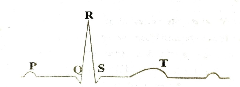
Q.28. What is ECG? What do p and T wave represent in a standard ECG.
Ans :- ECG is a graphical representation of the electrical activity of the heart during a cardiac cycle.
P-wave represent excitation of the atria and T-wave represent the return of the ventricles from excited to normal state.
Q.29. What is hepatic portal septem?
Ans :- A unique vascular connection exists between the digestive tract and liver.
Q.30. What is coronary system in our circulation?
Ans :- System of blood vessels present in our body exclusively for the circulation of blood to and from the cardiac musculature is termed us coronary system.
Q.31. What is pulmonary circulation?
Ans :- The deoxygenated blood pumped into the pulmonary artery is passed on to the lungs from where the oxygenated blood is carried by the pulmonary veins into the left atrium. This pathway constitute pulmonary circulation.
Q.32. What is system circulation?
Ans :- The oxygenated blood entering the aorta is carried by a network of arteries, carried by a network of arteries, arterioles and capillaries to the tissues from where the deoxygenated blood is collected by a system of venules, vines and vena cava & emptied into right atrium. This is the systemic circulation.
Q.33. What happens in atherosclerosis?
Ans :- It affects the vessels that supply blood to the heart muscle. It is caused by deposit of calcium, fat, cholesterol and fibrous tissues, which makes the lumen of arteries narrower..
Q.34. What are the major proteins present in plasma and also write their function.
Ans :- Major proteins are – Fibrinogen, globulin and Albumin.
Functions :-
Fibrinogen – help in blood clotting.
Globulin – involved in defence mechanism.
Albumin – help in osmotic balance.
Q.35. What role does the following cell plays in our body?
Neutrophil, Basophil and Eosinophil
Ans :- Neutrophil – Destroy foreign particles.
Basophil – involved in inflammatory reaction.
Eosinophil – resist infections and also associated with allergic reaction.
Q.36. Give the diagrammatic presentation of a standard ECG.
Ans :-
Q.37. How cardiac activity is regulated?
Ans :- Normal activity is regulated by nodal tissue. A special neural centre in medulla oblongata can mode rate the cardiac functions through autonomic nervous system. Neural signals through the sympathetic nerve can increase the rate of heart beat on other hand parasympathetic neural signals decrease the rate of heart beat, speed of conduction of action potential. Adrenal medullary hormones too can increase the cardiac output.
Q.38. Write the function of Lymph.
Ans :- Lymph is a colourless fluid rich in lymphocytes which are responsible for the immune responses of the body. It is also an important carrier for nutrient, hormones etc. Fats too are absorbed by lymph.
LONG ANSWER TYPE QUESTIONS
Q.39. Draw the internal structure of the heart and locate the following parts.
Ans :- Vena cava, tricuspid valve, semilunar valve, SAN, AVN, Purkinje fibre, pulmonary vein and pulmonary artery.

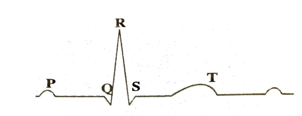
Q.40. Give diagrammatic presentation of a standard ECG. What does P ware and T ware represent?
Ans :- ECG is a graphical representation of the electrical activity of the heart during a cardiac cycle.
P-wave represent excitation of the atria and T-wave represent the return of the ventricles from excited to normal state.
Q.41. What is Rh grouping? Why is it necessary to check the Rh group in a pregnant women.
Ans :- In blood another antigen similar to one present in Rhesus monkey is also observed on the surface of RBCs of majority of human. This antigen is termed as Rh. The person having Rh is termed as Rh positive and the person not having Rh antigen are termed as Rh – ve.
A special case of Rh incompatibility has been observed between the Rh-ve blood of a pregnant mother with Rh+ve blood of the foetus. Rh antigens of the foetus do not get oxposed to the Rh-ve blood of the mother in the 1st pregnancy as the two blood are well separated by the placenta. However during delivery of the 1ˢᵗ child there is possibility of exposure of the maternal blood to small amounts of Rh+ve blood from the foetus. In much cases the mother start preparing antibodies oxygen Rh antigen in her blood. In case of her subsequent pregnancies the Rh antibodies (Rh-ve) from the mother can leak in to the blood of focus (Rh+ve) and destroy the foetal call. This could be fatal to focus or could cause severe anaemia and jaundice to the baby. So it is necessary to check the Rh group in a pregnant women.
Q.42. What is ABO grouping? Why ‘O’ blood group is called the universal donor and AB blood group the universal recipient?
Ans :- ABO blood grouping is based or the presence absence of two surface antigens (A and B) on RBCs. Also the plasma of different individuals contain 2 natural antibodies. In us 4 different types of blood group are there. They are A, B, AB and O group.
In blood group A, antigen A and antibody B are present.
In blood group B, antigen B and antibody B are present.
In blood group AB, both A and B antigens are present but it possess no antibodies and in blood group O there is no antigen …… It possess both A and B antibodies.
An individual with ‘O’ blood group can donate to person with any other blood group and hence ‘O’ group individual are called universal donors. Person with AB blood group can accept blood from persons with AB as well as the other group of blood. So such persons are called universal recipients.

Hi, I’m Dev Kirtonia, Founder & CEO of Dev Library. A website that provides all SCERT, NCERT 3 to 12, and BA, B.com, B.Sc, and Computer Science with Post Graduate Notes & Suggestions, Novel, eBooks, Biography, Quotes, Study Materials, and more.
